Craniosynostosis
Craniofacial Conditions > Craniosynostosis
What is Craniosynostosis?
Craniosynostosis is a birth defect in which one or more of the fibrous joints (sutures) between the bones of a baby’s skull close prematurely, before the brain has fully formed. This early closure can cause the skull to develop an abnormal shape as the brain continues to grow. Craniosynostosis can affect just one suture or multiple sutures, and the severity and symptoms vary based on the type and number of sutures involved.
Table of Contents
Craniosynostosis
Overview: Craniosynostosis is a condition where one or more sutures in a baby’s skull close too early, causing an abnormal head shape and potential issues with brain growth.
Prevalence: Craniosynostosis affects about 1 in every 2,000 to 2,500 births.
Causes: It can result from genetic mutations or occur spontaneously without a clear cause.
Diagnosis: Diagnosis is typically made through physical examination and confirmed with imaging studies like X-rays or CT scans, sometimes followed by genetic testing.
Common Characteristics: Features often include an abnormal skull shape, head or facial asymmetry, increased intracranial pressure, and sometimes developmental delays or vision issues.

Causes of Craniosynostosis
Craniosynostosis is typically a result of genetic factors, though in many cases, no specific cause can be identified. Some cases are associated with genetic syndromes, such as Crouzon, Pfeiffer, or Apert syndrome, where multiple sutures are affected, and other physical abnormalities may be present.
Types of Craniosynostosis
- Sagittal Craniosynostosis: The most common type, where the suture along the top of the skull closes early, resulting in a long, narrow head.
- Coronal Craniosynostosis: This occurs when one or both of the sutures running from ear to the top of the head close prematurely, leading to a flattened forehead and brow.
- Metopic Craniosynostosis: This affects the suture that runs from the nose to the top of the forehead, causing a triangular-shaped forehead.
- Lambdoid Craniosynostosis: Rare, this type involves the suture at the back of the head, potentially resulting in a flattened head shape.
Diagnosis
Craniosynostosis is often diagnosed soon after birth during a physical examination if an abnormal skull shape is observed. Further diagnostic tools include imaging studies, such as X-rays or CT scans, which provide detailed pictures of the sutures. In some cases, genetic testing may be recommended to check for associated syndromes.
Treatment
The treatment for craniosynostosis typically involves surgery to correct the shape of the skull and allow for proper brain growth. The type of surgery depends on the severity and type of craniosynostosis. In milder cases, surgery may be performed to relieve pressure on the brain and improve appearance, while more complex cases may require multiple surgeries to fully address the issue. The timing of surgery is critical, and most procedures are performed within the first year of life. Post-surgery, some children may need to wear a custom-fitted helmet to help reshape the skull.
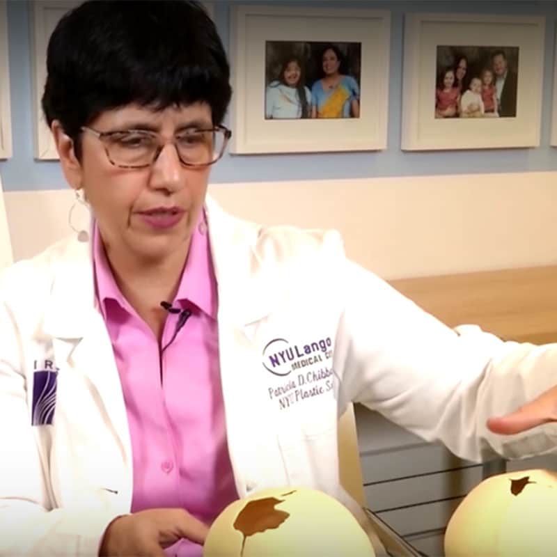
Surgery is the primary treatment for Craniosynostosis, aimed at correcting the premature fusion of skull sutures to allow the brain to grow properly and to improve the shape of the skull. The timing and type of surgery can depend on the severity of the condition and whether it’s syndromic or non-syndromic Craniosynostosis.
Surgery for Craniosynostosis
Specifically, procedures like Fronto-Orbital Advancement (FOA) and Le Fort III surgeries are designed to correct cranial and facial bone structures, which are key in managing craniosynostosis. These surgeries expand the space in the skull, which helps relieve brain compression and improve overall facial appearance, especially around the eyes. The Monobloc procedure is another relevant surgery for syndromic craniosynostosis, addressing both the skull and upper facial bones to enhance functionality and aesthetics.
Recovery:
- Fronto-Orbital Advancement (FOA):
- Typically requires a hospital stay of 3-5 days.
- Swelling and bruising are common in the first week, and it can take several weeks for the child to fully heal.
- Full recovery usually occurs within a few months, but follow-up appointments will be necessary to monitor skull growth.
- Children may need to wear a protective helmet to aid in the skull reshaping process.
- Le Fort III and Monobloc Surgeries:
- Recovery may take longer, with initial hospital stays ranging from 5-7 days.
- Swelling and discomfort can last for a few weeks, and sometimes, the child may require additional surgeries as they grow.
- Follow-up imaging and examinations will be scheduled to ensure proper healing and bone growth.
Risks:
- Infection: As with any surgery, there’s a risk of infection, particularly around incision sites.
- Blood Loss: These surgeries can involve significant blood loss, which may require transfusions.
- Swelling and Bruising: Facial and cranial swelling are common but usually subside within weeks.
- Neurological Complications: In rare cases, craniosynostosis surgeries can lead to nerve damage or neurological complications, including seizures or developmental delays.
- Recurrence: There is a slight risk that the sutures could prematurely fuse again, which may necessitate additional surgery.
Long-Term Outlook:
- Most children who undergo craniosynostosis surgery, particularly if performed early, go on to have normal brain development and good cosmetic outcomes.
- The risk of developmental delays, cognitive issues, and increased intracranial pressure is greatly reduced when treated early.
- Children with syndromic craniosynostosis, who often undergo Le Fort III or Monobloc surgeries, may require additional procedures as they grow due to ongoing facial and cranial changes.
- Long-term outcomes are generally positive, but they depend on the severity of the condition and whether it’s associated with other syndromes or complications. Regular follow-up care is critical to monitor growth and development.
What are the risks of not treating Craniosynostosis?
If Craniosynostosis is not treated, it can lead to significant complications. The premature fusion of the skull sutures limits brain growth, potentially causing increased intracranial pressure.
This pressure may result in:
- Developmental delays
- Cognitive impairments
- Vision issues.
In severe cases, untreated Craniosynostosis can cause seizures, hearing loss, and chronic headaches. Additionally, the abnormal shape of the skull can worsen over time, leading to social and psychological difficulties for the child. Early diagnosis and treatment are crucial for preventing these risks and ensuring healthy development.
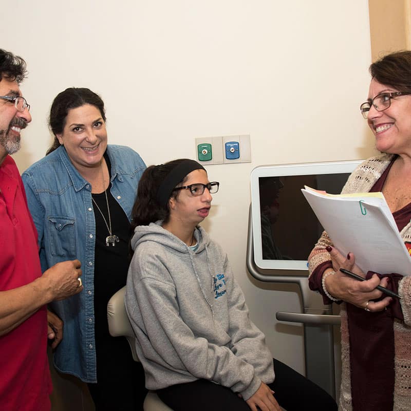
Explore Our Comprehensive Parent Guides
Supporting a child with a craniofacial condition can be challenging, but you don’t have to navigate it alone. Our expert-created Parent Guides offer valuable insights, practical tips, and resources to help you provide the best care for your child.
Living with Craniosynostosis
With early diagnosis and proper treatment, most children with craniosynostosis go on to lead healthy, normal lives. However, long-term follow-up with healthcare providers may be necessary to monitor skull growth, brain development, and any potential complications. Some children may experience developmental delays, vision problems, or require additional surgeries, depending on the severity of their condition.
Life Expectancy
The life expectancy for individuals with Craniosynostosis is generally normal, especially when the condition is diagnosed and treated early. Many cases are corrected through surgery, allowing for normal brain development and a healthy life. However, if left untreated, Craniosynostosis can lead to complications such as increased intracranial pressure, developmental delays, and neurological issues, which may impact overall quality of life.
In rare cases where Craniosynostosis is part of a syndrome involving other medical complications (such as in Apert or Crouzon syndrome), life expectancy can be influenced by the severity of associated health problems. Proper medical care and early intervention are key to managing the condition and ensuring a good prognosis.
Mild Craniosynostosis
Mild Craniosynostosis refers to cases where only one suture in the skull is prematurely fused, but the condition may not be as immediately noticeable or severe as more advanced forms. In these cases, the brain often has enough room to grow through the other, unfused sutures. As a result, symptoms like increased intracranial pressure or developmental delays might not appear, or they may be very mild.
Key Characteristics of Mild Craniosynostosis:
- Cosmetic Issues: The most common sign of mild craniosynostosis is an abnormal head shape, but without major functional concerns.
- Subtle Symptoms: Some children with mild forms may not show obvious symptoms early on, making diagnosis more challenging.
- Normal Brain Development: In many cases, cognitive development is normal, especially if the fusion is limited to a single suture.
- Delayed Diagnosis: Because symptoms are less severe, mild craniosynostosis may be diagnosed later in childhood or even adulthood.
Treatment for mild Craniosynostosis:
- Observation: In very mild cases, treatment might not be necessary, especially if brain development is unaffected. Doctors might opt for regular monitoring to ensure there are no complications.
- Minimally Invasive Surgery: If needed, endoscopic surgery may be recommended for cosmetic correction and to prevent future complications.
- Helmet Therapy: In some cases, helmet therapy might be used to gently reshape the skull as it grows, particularly for infants.
Resources and Support
Families dealing with craniosynostosis can benefit from connecting with support groups and specialists who understand the complexities of the condition. The following resources may be helpful:
- Craniosynostosis Support Groups: Online communities and local support groups where families can share experiences and advice.
- Specialists: Seeking care from a multidisciplinary team, including pediatric neurosurgeons, craniofacial surgeons, and genetic counselors, is essential for optimal outcomes.